I had a customer in Malaysia reach out asking about using the OpenBCI Cyton in a study they are running with mice. I was aware of one paper from 2023 that I shared with them, but as I looked around I found more than I expected!
Although our boards were not originally designed with animal research in mind, a growing number of researchers have managed to apply them that way. For example:
Abstract
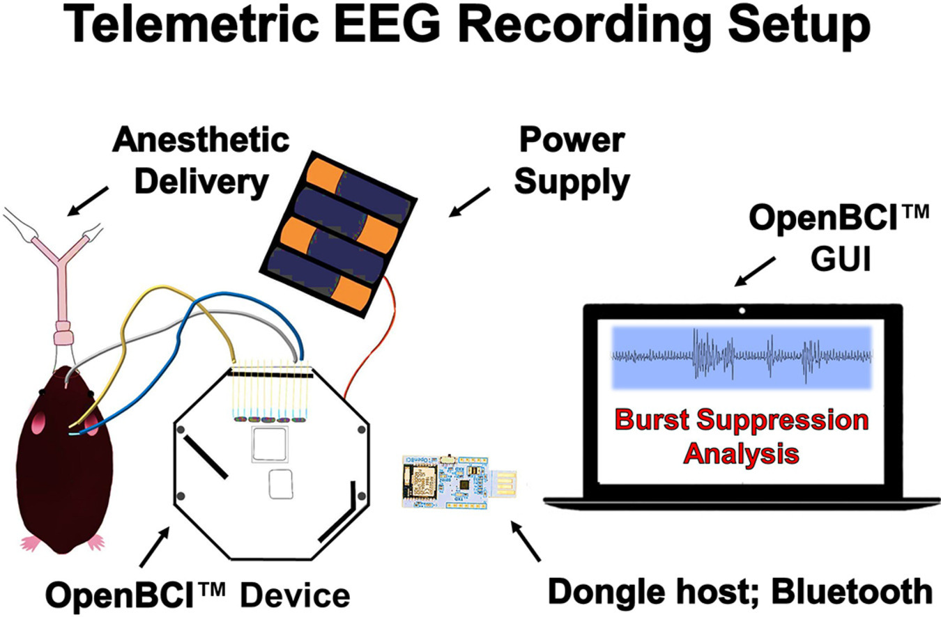
Telemetric electroencephalography (EEG) recording, using subdermal needle electrodes, is a minimally-invasive method to investigate mammalian neurophysiology during anesthesia. These inexpensive systems may streamline experiments examining global brain phenomena during surgical anesthesia or disease. We utilized the OpenBCI™ Cyton board with subdermal needle electrodes to extract EEG features in six C57BL/6J mice undergoing isoflurane anesthesia. Burst suppression ratio (BSR) and spectral features were compared for a verification of our method. Following an increase from 1.5% to 2.0% isoflurane, the BSR increased (Wilcoxon-signed-rank statistic; p = 0.0313). Furthermore, although the absolute EEG spectral power decreased, the relative spectral power remained comparable (Wilcoxon-Mann-Whitney U-Statistic; 95% CI exclusive AUC=0.5; p < 0.05). Compared to tethered systems, this method confers several improvements for anesthesia specific protocols: 1-Avoiding electrode implant surgical procedures, 2-Anatomical non-specificity for needle electrode placement to monitor global cortical activity representative of anesthetic state, 3-Facility to repeat recordings in the same animal, 4-User-friendly for non-experts, 5-Rapid set-up time, and 6-Lower costs. Minimally-invasive telemetric EEG recording systems ergonomically improve tethered systems for anesthesia protocols. Using this method, we verified that higher isoflurane concentrations resulted in an increased EEG burst suppression ratio and decreased EEG absolute spectral power, with no change in frequency distribution.
Mansouri, M. T., Ahmed, M. T., Cassim, T. Z., Kreuzer, M., Graves, M. C., Fenzl, T., & García, P. S. (2023). Telemetric electroencephalography recording in anesthetized mice—A novel system using minimally-invasive needle electrodes with a wireless OpenBCI™ Cyton Biosensing Board. MethodsX, 10, 102187. https://doi.org/10.1016/j.mex.2023.102187
An Improved Method for Electroencephalographic Detection of Epileptic Discharge
Abstract
Objective To investigate the role of OpenBCI module in the electroencephalographic (EEG) detection of epileptic discharge. Methods C57BL/6J mice aged 8-12 weeks were divided into two groups: the sham-operated group and kainic acid-induced epileptic group. Spontaneous seizures were monitored continuously for 3 weeks either by EEG or by OpenBCI. Results Up to 8 mice could be simultaneously monitored by OpenBCI. Meanwhile, the module accurately recorded the resting discharge, EEG spikes, and seizures. Conclusion Compared with the conventional brain function monitoring system, the OpenBCI module has lower cost and data occupancy and thus may be applied in clinical settings.
Shi XZ, Xu Q. [An Improved Method for Electroencephalographic Detection of Epileptic Discharge]. Zhongguo Yi Xue Ke Xue Yuan Xue Bao. 2019 Feb 28;41(1):53-56. Chinese. doi: 10.3881/j.issn.1000-503X.10418. PMID: 30837042.
A flexible implantable microelectrode array for recording electrocorticography signals from rodents
Abstract
Electrocorticography signals, the intracranial recording of electrical signatures of the brain, are recorded by non-penetrating planar electrode arrays placed on the cortical surface. Flexible electrode arrays minimize the tissue damage upon implantation. This work shows the design and development of a 32-channel flexible microelectrode array to record electrocorticography signals from the rat’s brain. The array was fabricated on a biocompatible flexible polyimide substrate. A titanium/gold layer was patterned as electrodes, and a thin polyimide layer was used for insulation. The fabricated microelectrode array was mounted on the exposed somatosensory cortex of the right hemisphere of a rat after craniotomy and incision of the dura. The signals were recorded using OpenBCI Cyton Daisy Biosensing Boards. The array faithfully recorded the baseline electrocorticography signals, the induced epileptic activities after applying a convulsant, and the recovered baseline signals after applying an antiepileptic drug. The signals recorded by such fabricated microelectrode array from anesthetized rats demonstrate its potential to monitor electrical signatures corresponding to epilepsy. Finally, the time–frequency analyses highlight the difference in spatiotemporal features of baseline and evoked epileptic discharges.
Chatterjee, S., Sakorikar, T., BS, A. et al. A flexible implantable microelectrode array for recording electrocorticography signals from rodents. Biomed Microdevices 24, 31 (2022). https://doi.org/10.1007/s10544-022-00632-0
Design and fabrication of a microelectrode array for studying epileptiform discharges from rodents
Abstract
Local field potentials, the extracellular electrical activities from brain regions, provide clinically relevant information about the status of neurophysiological conditions, including epilepsy. In this study, a 13-channel silicon-based single-shank microelectrode array (MEA) was designed and fabricated to record local field potentials (LFPs) from the different depths of a rat’s brain. A titanium/gold layer was patterned as electrodes on an oxidized silicon substrate, and silicon dioxide was deposited as a passivation layer. The fabricated array was implanted in the somatosensory cortex of the right hemisphere of an anesthetized rat. The developed MEA was interfaced with an OpenBCI Cyton Daisy Biosensing Board to acquire the local field potentials. The LFPs were acquired at three different neurophysiological conditions, including baseline signals, chemically-induced epileptiform discharges, and recovered baseline signals after anti-epileptic drug (AED) administration. Further, time-frequency analyses were performed on the acquired biopotentials to study the difference in spatiotemporal features. The processed signals and time-frequency analyses clearly distinguish between pre-convulsant and post-AED baselines and evoked epileptiform discharges.
Chatterjee, S., Joshi, R.K., Sakorikar, T. et al. Design and fabrication of a microelectrode array for studying epileptiform discharges from rodents. Biomed Microdevices 25, 31 (2023). https://doi.org/10.1007/s10544-023-00672-0
This one used the OpenBCI GUI software, combined with non-OpenBCI hardware and electrodes.
Abstract
Coronary heart disease (CHD) is a life-threatening disease caused by obstruction of the coronary arteries that interferes with blood flow known as atherosclerosis. Hyperlipidemia, a risk factor of atherosclerosis, is characterized by excessive concentrations of total cholesterol, LDL, and triglycerides with low concentrations of HDL. A high-fat diet (HFD) contributes to the progression of atherosclerosis, CHD, and other cardiovascular diseases. This study aims to measure electrocardiography (ECG) waves of hyperlipidemia-induced rats. Twenty rats were fed different diets for eight weeks, i.e., the control group (normal diet) and the HFD group (high-fat diet). Their ECG was recorded using a Wireless Mice Electrocardiogram (WIM ECG) for 5-10 min. After eight weeks, the HFD group showed a significantly
higher lipid profile concentration (cholesterol: 179.03 mg/dL, triglyceride: 149.11 mg/dL, LDL: 123 mg/dL, HDL: 29.15 mg/dL) than the control. This hyperlipidemic condition causes a significant change in some characteristics of the ECG wave. At week 8, the characteristic ECG wave duration for the HFD groups was RR intervals (176.5 ms), QT intervals (123.5 ms), T waves (33.6 ms), P wave (27.4 ms), QRS interval (64.9 ms), ST-segment (23.7 ms), and heart rate (334 bpm). This study concludes that long-period HFD feeding in rats leads to hyperlipidemia and causes changes in the characteristics of ECG waves.
Maulana, H., Ridwan, A., Suprijanto, S., Shanty Rahayu Kusumawardani, S. R. K., & Fitri, L. L. (2023). Electrocardiogram Analysis of Wistar Rats Induced by Hyperlipidemia using Wireless Mice Electrocardiogram (Wim ECG) in Coronary Heart Disease Study. Sains Malaysiana, 52(2), 589–597. https://doi.org/10.17576/jsm-2023-5202-20
Most of this research used either our Cyton or Cyton+Daisy bioamplifier.
For more research from the OpenBCI community, check out the Citations Page https://openbci.com/citations
Have you done animal research using OpenBCI? Let us know in the comments or at [email protected]
Leave a Reply
You must be logged in to post a comment.